Kidney Stone Treatment & Diagnosis
Kidney Stone Diseases
Diagnosis and treatment for kidney, ureter, bladder, and urethral stones, from non-surgical options to PCNL and laser lithotripsy.
Diagnosis & Treatment for Kidney Stone Diseases
Our Division of Stone Disease treats stones anywhere they form in the urinary tract, kidneys, ureters, bladder, or urethra. The large majority, around 97%, form in the kidneys and ureters, with the remaining 3% occurring in the bladder or urethra.
Not every stone needs the same approach. Treatment depends on where a stone is, how large it is, and whether it's causing symptoms, which is why we start with a proper diagnostic work-up before recommending a path forward.
Problems Treated
Characteristics of Stones
- Composition: Urinary stones form from a mix of mineral and organic material. Calcium oxalate is by far the most common type, making up roughly 80% of all stones.
- How Common They Are: Around 15% of people will develop a kidney stone at some point in their life. It's rarely life-threatening, but it can cause severe pain, infection, and, if left untreated, kidney damage. Even with modern treatment, stones tend to recur, largely because we still don't fully understand why they form in some people and not others.
Formation of Stones
Stone formation is influenced by a mix of genetics, diet, certain medications, and anatomical factors. In simple terms, a stone forms when certain substances in urine become concentrated enough to crystallize, a process made worse when the body lacks the natural inhibitors that normally keep crystals from growing.
Clinical Features
Symptoms vary depending on the stone's size and location, but commonly include:
- Pain ranging from mild discomfort to severe episodes
- Blood in the urine
- Passage of small tissue fragments in the urine
- Urinary tract infections
- A burning sensation during urination
- Urinary blockage
- No symptoms at all, some stones are only found incidentally on imaging
Treatment of Stones
The first priority is always pain relief. From there, treatment focuses on helping the stone pass or, if that's unlikely, removing it directly.
Most small stones pass on their own: roughly 80-90% of stones under 5 mm, and even some smooth stones up to 7-8 mm, will clear without intervention. Larger stones typically need active removal, and if there's an underlying anatomical issue contributing to stone formation, we address that at the same time.
For a single kidney stone between 1-2 cm, ESWL (Extracorporeal Shock Wave Lithotripsy) is often the first choice. It uses focused shock waves from outside the body to break the stone into pieces small enough to pass naturally, usually over the following 1-12 weeks.
Larger kidney stones are generally better suited to PCNL (Percutaneous Nephrolithotomy), a procedure that accesses the kidney through a small puncture in the back to remove the stone directly, we cover this in more detail below.
When a larger stone is sitting in the lower ureter, or hasn't passed after about a month, laser lithotripsy is typically used: a thin endoscope is passed into the urinary tract and a laser breaks the stone apart from the inside.
Open surgery is rarely needed for ureteric stones today and is reserved for unusually complex cases.
Uric acid stones, which often don't show up on X-ray and are usually spotted on ultrasound instead, can sometimes be dissolved without surgery by alkalinizing the urine, provided they're smaller than about 1 cm.
Diet plays a real role in preventing future stones. Depending on your stone type, we typically recommend:
- More neutral fluids, water, tender coconut water, diluted buttermilk, citrus juices, while keeping coffee, tea, and milk to around 1-2 cups a day.
- A fiber-rich, largely vegetarian diet where possible.
- For meat eaters, keeping protein intake to around 1 gram per kilogram of body weight per day.
- Cutting back on sugary and soft drinks, and avoiding calcium-rich foods on an empty stomach.
Diagnostic Tests
We start most patients with our Stone Pack (SP) tests, a set of blood and urine tests that look at stone-formation risk, blood sugar, kidney function, and general blood health.
Imaging typically includes an abdominal ultrasound and an X-ray KUB (kidney, ureter, and bladder) to locate stones. A CT scan of the KUB region is the single most sensitive test we have for detecting stones and is often used when other imaging isn't conclusive.
Depending on the situation, your urologist may also order additional functional tests, intravenous urography, contrast CT KUB, or an isotope renogram, to better understand how well the kidneys are functioning.
For patients with recurrent stones, a family history of stone disease, or other complicating factors, we offer more detailed testing, including SP3 (fasting and random blood tests) and SP4 (a 24-hour urine collection repeated over 3 consecutive days).
Any stone that's removed or passed can be analyzed using FTIR (Fourier-transform infrared spectroscopy), one of the most precise ways to identify exactly what a stone is made of. That result often shapes the specific dietary and medication recommendations your doctor gives you afterward.
Percutaneous Nephrolithotomy (PCNL)
PCNL is a minimally invasive procedure for removing larger kidney or upper ureter stones, offering a notably easier recovery than traditional open surgery.
How PCNL Is Performed
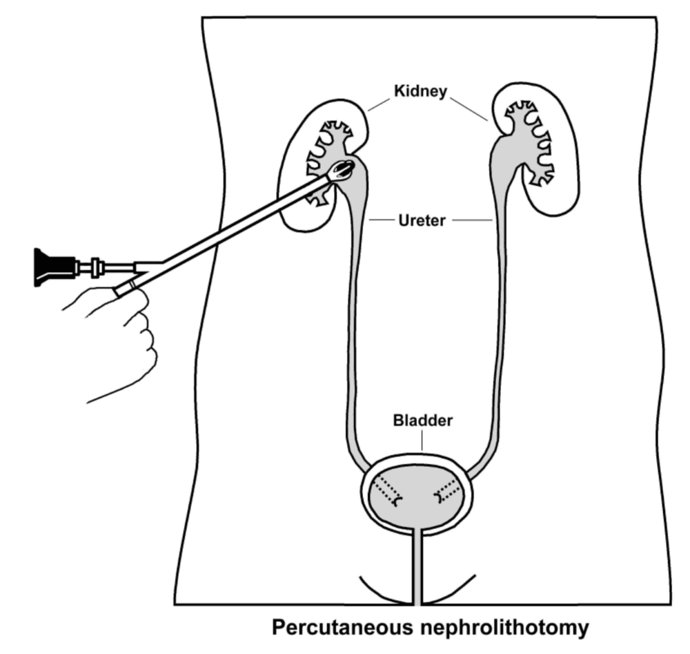
The surgeon makes a small incision, typically 1 cm or less, in the flank, then guides a wire into the kidney using X-ray (fluoroscopic) guidance. The passage is gradually widened so a nephroscope can be inserted to see and remove the stone directly. Larger stones are broken into smaller pieces first to make removal easier. A drainage tube usually stays in place for 1-2 days afterward to support healing.
What sets PCNL apart is that it removes the stone in a single sitting under direct visualization, unlike ESWL or ureteroscopy, which sometimes need repeat sessions, and unlike open surgery, which requires a much larger incision. Most patients spend around 3-4 days in the hospital.
We typically recommend PCNL for kidney stones larger than 2 cm or upper ureteric stones larger than 1 cm that aren't good candidates for ureteroscopy or ESWL, as well as for stones that haven't responded to other treatments.
The procedure is usually done under general anesthesia, though intravenous sedation, regional, or local anesthesia may be used instead depending on the patient's overall health.
Stone clearance rates with PCNL are generally between 90-95%; a small number of patients need a second procedure depending on stone size, number, and location.
After surgery, most patients stay in hospital for 2-3 days, with follow-up imaging (X-ray or ultrasound) to check for any remaining fragments. If needed, a second PCNL or a round of ESWL can clear what's left.
A drainage tube and urinary catheter are usually kept in for 1-2 days. Some brief urine leakage around the puncture site is common for a day or so after the tube comes out, we'll ask you to drink enough fluid to keep urine output above roughly 2 litres a day during recovery.
As with any surgery, there are risks. Bleeding significant enough to need a transfusion occurs in about 1% of cases, and infection is possible. Ongoing urine leakage or fever after the procedure should be reported promptly, as these can signal a complication needing further treatment.
Rarely, more serious complications occur, including uncontrolled bleeding (roughly 0.4% of cases) or a collapsed lung (pneumothorax). These are uncommon but are managed with further intervention when they do happen.
Most people return to normal daily activities within about a week, though we recommend holding off on strenuous activity for at least two weeks. Full recovery time depends on how much stone burden was present and how many access points were needed during surgery.
PCNL isn't suitable for everyone, it's generally avoided in patients with bleeding disorders, during pregnancy (due to radiation exposure), or where a patient's overall health makes anesthesia too risky. Afterward, we often recommend dietary changes, generally a higher-fiber diet and reduced intake of high-calorie foods, alongside any medications suited to what's actually driving your stone formation.