Paediatric Urology Treatment & Diagnosis
Paediatric Urology Care for Children
Diagnosis and treatment for urological conditions in children, from UTIs and voiding disorders to hypospadias, VUR, and reconstructive surgery.
Treatments & Diagnosis for Paediatric Urology
Our Paediatric Urology team treats urological conditions in children, from routine issues like undescended testicles to more complex reconstructive surgery for conditions like bladder exstrophy. Children have different anatomy, physiology, and emotional needs than adults, so treatment is built around that rather than adapted from adult urology.
Who is a Paediatric Urologist?
If your child has an illness or disease of the genitals or urinary tract (kidneys, ureters, bladder), a Paediatric Urologist has the experience and qualifications to treat your child.
Paediatric Urologist
Children are not small adults. They cannot always say what is bothering them. They cannot always answer medical questions and are not always able to be patient and cooperative during a medical examination.
Paediatric Urologists are specially trained to focus on the unique medical needs of children. They use gentle techniques to examine and treat children, making the experience comfortable and non-threatening. This includes using specialized equipment tailored for children to ensure their well-being during the process.
If your Paediatrician recommends a Paediatric Urologist, that referral reflects a level of specialisation your child's regular doctor doesn't have, specifically in children's urinary tract and genital health.
Paediatric Urologists Training
Paediatric Urologists are medical professionals who undergo extensive training:
- At least 6 years of medical school
- 1 to 3 years of surgical residency
- At least 3 additional years in general Urology Health
- 1 to 2 years of specialized fellowship training in Paediatric Urology
They typically dedicate the large majority of their practice specifically to treating urinary and genital problems in children, rather than splitting time with general adult urology.
Paediatric Urologists Treatment
Paediatric Urologists specialize in diagnosing, treating, and managing a wide range of urinary and genital issues in children. Their treatments include:
- Management of voiding disorders, vesicoureteral reflux, and urinary tract infections that may require surgery
- Surgical reconstruction of the urinary tract, including correction of genital abnormalities and disorders of sex development
- Treatment of groin conditions such as undescended testes, hydrocele/hernia, and varicocele
- Kidney stone treatment and surgical management
- Addressing tumours and malignancies of the kidney, bladder, and testis
- Management of urological problems identified before birth
- Treatment of urinary tract problems linked to neurological conditions like spina bifida
Antenatal/Fetal Hydronephrosis
Antenatal Hydronephrosis refers to the fluid-filled enlargement of the kidney before birth, detected via ultrasound. While most cases do not affect obstetric care, some may require surgery during infancy or childhood. Management during pregnancy usually involves regular ultrasound surveillance.
Common Causes of Antenatal Hydronephrosis:
Blockages: These may occur at various points in the urinary system, such as the Uretero Pelvic Junction (UPJ) or the Uretero Vesical Junction (UVJ).
Reflux: Vesicoureteral reflux causes urine to flow back into the kidney, potentially requiring antibiotic prophylaxis and regular monitoring.
Other Conditions: Duplicated kidneys or multicystic kidneys may show up during ultrasound and require follow-up care.
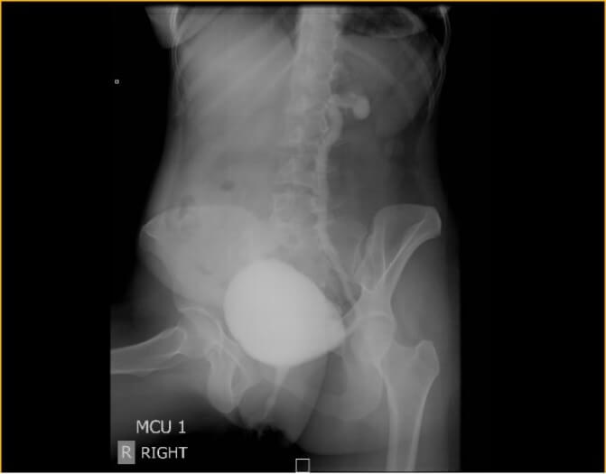
Management After Birth: Postnatal ultrasound and specialized imaging such as Voiding Cystourethrogram (VCUG) are typically used to evaluate and treat the condition.
Circumcision/Circumcision Revision
Circumcision is a common procedure, either done routinely or due to medical reasons such as infections or irritation. It may need to be performed or revised in young children under general anesthesia, correcting adhesions or excess skin.
Postoperative Care: After circumcision, it's important to apply antibiotic ointment or Vaseline to protect the exposed glans during diaper changes, especially during the healing period.
For circumcision revisions, waiting until the child is 6 months old helps minimize cosmetic risks while ensuring the procedure is effective and safe.
Hypospadias
Hypospadias is a congenital condition in boys where the urethral opening is located on the undersurface of the penis instead of the tip. It can range from mild to severe, and in some cases, is accompanied by chordee (penile curvature). Hypospadias affects roughly 8 per 1,000 male births, and there is a hereditary component, with around a 15% chance of recurrence in family members.
The condition is classified based on the location of the urethral opening, from near the glans penis to the perineum (anal opening). The presence of chordee affects the type of surgery required, and the correction may involve one or two stages depending on the severity.
Why Correct Hypospadias?
Surgical correction involves straightening any chordee and extending the urethra to the tip of the penis, which matters for normal urinary and sexual function in adulthood, even though the need for correction is identified in early childhood.
Hypospadias and Chordee Surgery
Several surgical techniques are used to treat hypospadias, with advancements reducing the number of surgeries needed. Most children undergo the procedure between 6 to 8 months of age, and typically, a single surgery is sufficient. Severe cases may require two operations. The surgery is outpatient and performed under general anesthesia. After surgery, a catheter may be needed for one to two weeks for drainage.
Potential Complications
Infection and bladder spasms are uncommon but possible. Fistulas (leaks in the new urethra) occur in about 10% of more complex surgeries and may require a follow-up procedure. Strictures (narrowing due to scar tissue) may also develop at the new urethra opening.
Labial Adhesions
Labial adhesions occur when the inner folds of the female genitalia (labia) temporarily stick together, primarily in young girls aged 3 months to 6 years. Hormonal factors and skin inflammation contribute to the condition, often linked to irritation or minor infection. Although labial adhesions are mostly asymptomatic, post-void dribbling may occur, and urinary tract infections are rare.
Initial treatment involves the application of estrogen cream twice daily for 2-8 weeks. In some cases, a doctor may separate the adhesions using topical anesthetic or general anesthesia. After the adhesions resolve, it's important to apply a thin layer of estrogen cream nightly to prevent recurrence.
Neurogenic Bladder
What is Neurogenic Bladder (NB)?
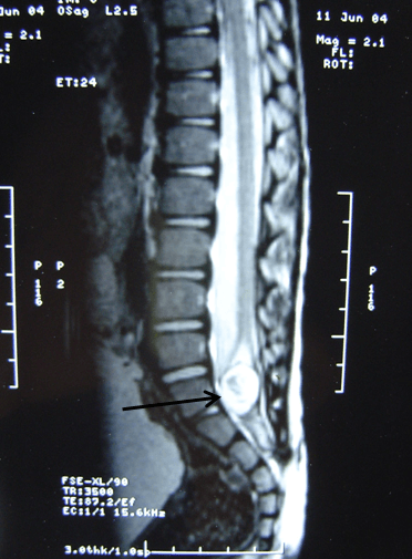
Neurogenic Bladder (NB) refers to a dysfunctional lower urinary tract caused by issues other than primary urinary tract pathology. It occurs when neurological conditions impact the bladder's control.
Normal Urination Process
Under typical circumstances, urination is voluntary, initiated by the brain and controlled through the spinal cord and nerves. Coordination between the bladder and sphincters ensures proper urine flow. Disruptions in this process, such as involuntary leakage or difficulty urinating, require medical attention.
The control of urination involves messages from the brain traveling through the spinal cord to the bladder. Any damage to the spinal cord or nerves can lead to the loss of bladder control, resulting in either urinary retention or leakage.
Conditions That Cause Neurogenic Bladder
Conditions like Parkinson's disease, Alzheimer's, multiple sclerosis, spinal cord injury, diabetes, and others can lead to neurogenic bladder by affecting the brain or spinal cord.
Symptoms
Patients with neurogenic bladder may experience urinary retention, incontinence, frequency, urgency, loss of bladder sensation, and difficulty controlling bowel movements.
Bladder dysfunction itself, beyond the visible physical disability from a neurological injury, is a significant contributor to renal failure and illness if left untreated, which is why it needs its own dedicated evaluation.
Investigations for Diagnosis
Diagnosis includes tests such as urine analysis, ultrasound, MCU (micturating cystourethrogram), and urodynamic studies to assess bladder pressures and sphincter activity. MRI scans of the spine or brain help identify underlying causes in the nervous system.
How is Neurogenic Bladder Treated?
Treatment focuses on preventing kidney damage caused by increased bladder pressure. Measures aim to relieve bladder pressure, prevent infections, and manage urinary incontinence.
Treatment Options:
- Self-catheterization to empty the bladder regularly and prevent incontinence.
- Medications to reduce bladder pressure, often taken long-term.
- Surgical interventions, such as adding bowel to increase bladder capacity or inserting an artificial sphincter.
Bowel management is crucial and may include dietary changes, enemas, or laxatives.
Pelviureteric Junction (PUJ) Obstruction
What is Pelviureteric Junction (PUJ) Obstruction?
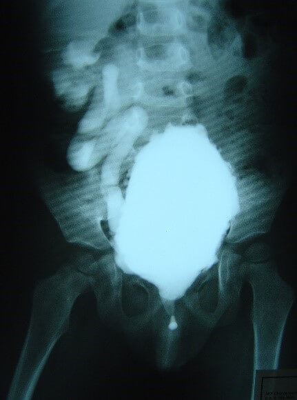
PUJ obstruction is a blockage between the renal pelvis and ureter, leading to impaired urine flow and potentially causing kidney damage if untreated. This condition can lead to hydronephrosis, a swelling of the kidney.
Causes of PUJ Obstruction
PUJ obstruction is commonly congenital due to abnormal muscle development, but can also result from blood vessel compression, inflammation, or scar tissue.
Symptoms of PUJ Obstruction
Symptoms include hematuria, urinary tract infections, kidney stones, abdominal pain, and hypertension. In infants, PUJ obstruction is often asymptomatic, detected through prenatal ultrasounds.
Diagnostic Tests for PUJ Obstruction
Confirmatory tests include ultrasound, nuclear scans (DTPA or EC renogram), and urine tests to assess kidney function. CT scans are avoided due to high radiation exposure.
Does PUJ Obstruction Require Surgery?
Surgery is recommended when symptoms or kidney function are impaired. The primary goal of surgery is to restore normal urinary flow and prevent kidney damage.
Surgical Treatment: Pyeloplasty
Pyeloplasty involves removing the scar tissue blocking the PUJ and reconnecting the ureter to the renal pelvis. In children, open surgery is typically performed, while adults may undergo laparoscopic surgery.
A ureteric stent is often placed during surgery to aid drainage. Rarely, a nephrostomy tube is used to drain the kidney if the stent cannot be placed.
Post-Surgery Care
Post-surgery care includes managing diet, monitoring for pain, and removing drainage tubes as needed. Follow-up ultrasounds and nuclear scans monitor recovery and kidney function.
Complications if Left Untreated
If untreated, PUJ obstruction can lead to kidney function loss, stone formation, and infection.
Follow-Up After Surgery
Renal ultrasounds and nuclear scans are performed periodically to ensure proper kidney function and drainage after pyeloplasty.
Successful Surgery Indicators
Success is measured by relief of symptoms, reduced hydronephrosis, and improved kidney function after surgery.
Testicular Self-Examination
Regular testicular examination should be part of every adult male's health routine, particularly once past adolescence. Testicular cancer is most common in men aged 20 to 40. Self-examination helps detect abnormalities early. By learning the normal anatomy of the testicles, men can spot changes that might signal potential issues.
It's best to perform the exam in a comfortable setting, such as the shower or bathtub. Examine the shape and position of the testicles, noting a groove called the lateral sulcus on the lateral surface, between the testicle and the epididymis. The testicle should feel firm, but not overly hard.
If testicular pain occurs, it may signal a torsion that requires surgery to prevent testicle loss. Early operation within 4-6 hours can save the testicle by restoring blood flow. The testicle is fixed to the scrotal wall to prevent recurrence. Consult a urologist immediately for acute testicular pain, don't wait to see if it resolves.
Torsion of the Testicle
Testicular torsion is a pediatric urologic emergency caused by the twisting of the blood vessels supplying the testicle. This affects the blood flow, leading to ischemia and potentially permanent testicle damage if left untreated.
Symptoms include sudden, severe scrotal pain, often accompanied by nausea and vomiting. It can occur during physical activity, accidents, or even at rest. Diagnosis can be challenging, but acute scrotal pain should be treated as torsion until proven otherwise.
If torsion is detected early (within six hours), the testicle can usually be saved. Delayed treatment increases the risk of infertility. Immediate detorsion and surgical fixation are required. The unaffected testicle is also secured to prevent future torsion.
Intermittent torsion may cause pain that resolves, but the risk of permanent damage remains. Elective fixation is recommended to prevent recurrence and protect testicular health.
Urinary Frequency
Urinary frequency is a common issue in pediatric urology. The condition is characterized by frequent urges to urinate, often with little urine output. It typically starts around age 5 and can be caused by increased bladder sensation.
Evaluation begins with a urinalysis to rule out infection or diabetes. Most children can control their bladder but may experience frequent urination, even at night. Reassurance and gradual improvement over time are key components of treatment.
In some cases, medications that address bladder sensitivity may help reduce symptoms. However, most children improve with time and decreased focus on bladder sensations.
Urinary Tract Infection in Children
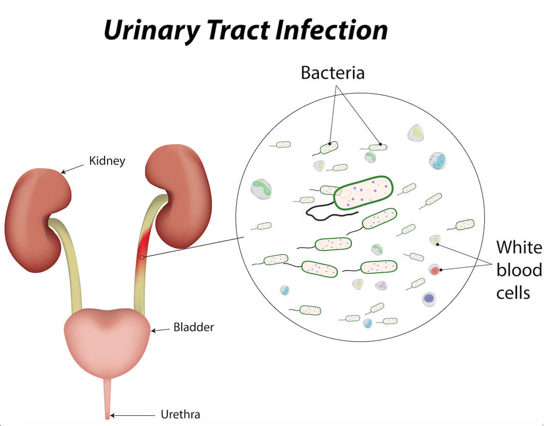
Urinary Tract Infection (UTI) in children is common but often undiagnosed. UTIs can occur in both healthy children and those with urinary tract abnormalities. Early detection is critical, as infants with UTIs may not show typical symptoms, making timely diagnosis essential for effective management.
How common is the problem?
Approximately 6% of girls and 2% of boys will experience a UTI during childhood.
What are the symptoms of UTI in children?
Unlike adults, children may not show typical UTI symptoms like burning or blood in urine. Infants may only show fever, poor feeding, or vomiting, making it easy to miss the diagnosis without suspicion.
What causes UTI in children?
UTIs in children often result from bacteria ascending from the genital area, with girls being more prone due to their shorter urethra. Other factors like urine flow blockage or reflux can also contribute to UTIs.
How is UTI diagnosed in children?
A simple urine test is essential when a child has unexplained fever. If a UTI is detected, further tests like urine culture, ultrasound, or imaging studies may be required to identify any underlying urinary tract issues.
How is UTI treated in children?
UTIs are treated with antibiotics, with the specific medication chosen based on urine culture results. Treatment may last up to 2 weeks, depending on the severity.
Can UTI be prevented?
Preventive measures include proper fluid intake, frequent bladder emptying, and a high-fiber diet to prevent constipation, which can exacerbate UTIs.
Does UTI have long-term effects?
Untreated UTIs can cause kidney damage and scarring, leading to long-term issues such as high blood pressure and reduced kidney function. Early detection and treatment are vital to prevent serious complications like kidney failure.
Urinary Tract Infection in Children
Voiding Disorders in Children
Voiding dysfunction in children, such as frequent urination, urgency, and wetting, is common and can cause concern for parents. These symptoms are often related to an immature bladder, especially in younger children.
- Frequent urination (frequency)
- Urgency to urinate (urgency)
- Wetting (incontinence, enuresis)
For young children, bladder maturation is key to successful toilet training. By age 4, most children achieve adult-like bladder control, but some may exhibit voiding issues as their bladder matures.
While some transient voiding symptoms are normal, persistent issues after toilet training require further evaluation to rule out anatomical or neurological problems.
Diagnosis and Treatment
Initial evaluation typically includes a physical exam and urine test. If necessary, imaging studies like ultrasound or voiding cystogram can help diagnose underlying conditions. Treatment may involve bladder training, timed voiding, or medications such as anticholinergics to address urgency and frequency.
Managing Voiding Disorders
In cases of persistent daytime symptoms, children may benefit from a structured voiding schedule or medications. For nighttime wetting, treatments like bed alarms or vasopressin may be effective.
In cases of severe or refractory symptoms, further tests such as urodynamic studies may be needed to determine the best course of action. With proper treatment, most children will eventually outgrow these voiding issues.
Meatal Stenosis Treatment
Meatal Stenosis is the abnormal narrowing of the urethral opening (meatus), which can make urination difficult and may prevent the bladder from emptying completely.
This condition is commonly found in circumcised boys. One theory suggests that the narrowing is caused by chronic exposure to urine irritation and the friction of the meatus against clothing or diapers, leading to inflammation and eventual scarring.
Symptoms of meatal stenosis include:
- Painful urination
- Straining to urinate
- Frequent urination
- Decreased caliber (thin) urinary stream
- Spraying while urinating
- Upward deflection of the urine stream
- Blood in the urine
Treatment for meatal stenosis can be addressed with one of two surgical methods. In meatoplasty, the meatus is widened by making an incision in the urethral opening, followed by the placement of a vascularized flap (often from the prepuce or surrounding penile skin) to prevent recurrence. In meatotomy, a small incision is made in the meatus, and the cut margins are either sutured with dissolvable stitches or left to heal naturally. Your surgeon will discuss the ideal treatment plan based on your child's specific condition. Some children may require additional dilation of the urethral opening post-procedure to prevent further narrowing.
Bladder Augmentation & Substitution
Main Reasons for These Operations
Conditions like Spina Bifida, posterior urethral valves, or bladder exstrophy may cause bladder dysfunction, leading to high urine pressure and kidney damage.
For patients with small bladders, bladder augmentation is needed to relieve symptoms and protect kidney function. In severe cases, bladder removal and substitution are required, particularly for bladder cancer or pelvic organ cancer. Surgery involves using intestine to reconstruct a normal-appearing bladder. Post-surgery, bowel rest and tube feeding may be necessary.
Potential Complications of Bladder Augmentation and Substitution
Catheterization: Patients need intermittent catheterization to empty the newly constructed bladder, as the intestine doesn't function like the normal bladder.
Mucus Formation: Intestinal mucus can obstruct catheters and lead to bladder stones or infections. Regular irrigation and long-term mucus evacuation are essential.
Intestinal Complications: Rare but serious complications like intestinal leakage or bowel obstruction may occur post-surgery. Prompt treatment is required for these risks.
Bladder Rupture/Perforation: A major risk after surgery is bladder rupture, usually due to inadequate catheterization. Symptoms include abdominal pain, fever, and infection.
Bladder Cancer: Patients on intermittent catheterization or those who've had bladder augmentation have an increased risk of bladder cancer.
Constipation
Under the Rome IV criteria, functional constipation in children is diagnosed when at least two of the following symptoms persist for at least one month:
- Fewer than three bowel movements per week
- Straining in over 25% of defecation attempts
- Lumpy or hard stools in over 25% of defecation attempts
- Sensation of anorectal obstruction or blockage
- Sensation of incomplete defecation
- Manual maneuvering required to defecate
Constipation is common among children with urinary tract infections and wetting issues. Treatment includes regular bowel habits, proper toilet training, and dietary changes.
Dietary Recommendations for Constipation Relief:
Increase intake of fruits (e.g., prune, pear, apricot) and vegetables (eat the peels) to help improve bowel movements. Bran cereal and bananas are also beneficial.
Foods that may worsen constipation:
Dairy products, rice, white bread, and potatoes can exacerbate constipation.
Medical Treatment: In some cases, laxatives such as Miralax or mineral oil may be prescribed to soften stool. Regular enemas may also be needed for children with neurological conditions.
Kidney Stones
Kidney stones are solid deposits formed from crystallized substances in the urine. They range in size from small grains to large staghorn stones, which may fill the entire kidney.
While small stones may pass unnoticed, larger stones can cause severe pain, urinary obstruction, and blood in the urine. Causes of kidney stones include dehydration, diet, infections, and genetic factors.
Symptoms of Kidney Stones:
- Severe back or flank pain
- Blood in the urine
- Fever and chills (infection)
- Vomiting
- Frequent or painful urination
Treatment depends on the stone's size and location. Options include shock wave therapy (ESWL), endoscopy (URS), and surgery (PCNL or Pyelolithotomy). Prevention involves increased water intake and dietary adjustments, with medication sometimes necessary to prevent recurrence.
Multicystic Dysplastic Kidney
Multicystic Dysplastic Kidney (MCDK) is a congenital kidney condition where the kidney is replaced by multiple cysts, resembling a bunch of grapes. The condition often presents with a non-functioning kidney, which is frequently diagnosed through prenatal ultrasound. Affected kidneys may not communicate with the ureter, and in some cases, the ureter may be absent. MCDK is more common in males, with a slight left-sided predominance. Close to half of cases are detected prenatally, with a smaller share presenting later as a flank mass.
The diagnostic process includes a renal ultrasound that distinguishes MCDK from hydronephrosis, a condition that mimics MCDK. Additional tests like DMSA or MAG-3 renal scans confirm the lack of kidney function. A contralateral kidney abnormality is found in a meaningful minority of cases, and conditions like vesicoureteral reflux may also be present. When MCDK is detected prenatally, assessing the opposite kidney and amniotic fluid volume is crucial to determine the viability of the baby.
The treatment options for MCDK are debated. While some recommend surgical removal of the affected kidney, others opt for observation. Although the risk of hypertension and renal tumors is low, yearly monitoring with renal ultrasound is recommended if the kidney is not removed. Tumors have been reported, both in childhood and adulthood, but they are extremely rare.
In rare instances, the diagnosis of MCDK remains uncertain despite imaging studies, and surgical exploration may be necessary for clarification.
Sleep Enuresis in Children
Sleep Enuresis, also known as bed-wetting or nocturnal enuresis, is a common condition affecting an estimated 10-20% of children around 5 to 6 years old. It is more prevalent in boys, and the condition often has a familial link. Children with a family history of enuresis are more likely to experience this issue. Primary enuresis occurs when the child has been bed-wetting since birth, while secondary enuresis starts after toilet training, between the ages of 5 and 8 years.
Enuresis is often classified as monosymptomatic nocturnal enuresis (MNE) when it occurs only at night, without daytime symptoms like urgency or frequency. The condition generally resolves with age, and many children experience spontaneous improvement, with only a small percentage of teenagers still affected by age 15. MNE is not typically linked to psychiatric or neurological disorders and often reflects developmental delay.
The treatment for MNE involves a combination of behavioral modifications and drug therapy. Children under 7 years old should not undergo treatment due to poor success rates. Medications like Desmopressin (DDAVP) may help reduce nocturnal urine production, but relapse rates are high once treatment is stopped.
Behavioral therapy, such as bladder training and the use of urinary alarms, is often effective. Parents can also support the child by reducing liquid intake in the evening, avoiding caffeine, ensuring adequate sleep, and using positive reinforcement for dry nights. A thorough evaluation of the child's urinary system is necessary to rule out other conditions, such as urinary tract abnormalities or bladder overactivity.
If bed-wetting persists or is accompanied by daytime symptoms, further diagnostic tests, including ultrasound and imaging, may be required to identify potential causes.
Spina Bifida Treatment
Children born with spina bifida often experience urinary tract abnormalities. The urinary tract includes the kidneys, ureters, bladder, and urethra, working together to filter waste and remove urine.
The kidneys filter waste from the blood and produce urine, which drains through the ureters into the bladder. The bladder stores urine, and the urethra releases it through urination. Proper bladder function relies on the sacral spinal cord's integrity to signal fullness and allow control over urination.
Many children with spina bifida have damaged nerves affecting bladder function, leading to poor urinary control and potential kidney and bladder damage. Immediate evaluation includes renal ultrasound and voiding cystourethrogram (VCUG) to assess kidney and bladder health.
Urodynamic studies are essential to assess bladder pressure and determine risks of kidney damage. These tests measure bladder pressure and can predict whether high pressures will lead to kidney harm, with over 35 cm H20 being concerning.
Managing Neurogenic Bladder:
Low Leak Point Pressure (<35 cm H20) - Children with low bladder pressures can be monitored regularly with annual renal ultrasounds and urodynamic follow-ups.
High Leak Point Pressure (>35 cm H20) - Children with high bladder pressures may require intermittent catheterization and medications to manage bladder function and prevent kidney damage.
Spontaneous Voiding: A small percentage of children with spina bifida can void spontaneously but need ongoing monitoring for potential spinal cord tethering and bladder function loss.
Urinary Tract Infections: Common in children with spina bifida, UTIs occur more frequently in those with high bladder pressures. Preventative care includes frequent bladder emptying and maintaining low bladder pressure.
Incontinence Management:
1. Surgical Options for High Bladder Pressure: Temporary procedures like vesicostomy can help reduce bladder pressure and facilitate proper urine drainage.
2. Medical Management: Anticholinergic medications and other treatments help relax bladder muscles and manage incontinence, although side effects like dry mouth or constipation should be monitored.
3. Surgical Interventions for Incontinence: In severe cases, bladder enlargement or sphincter surgery may be needed if medications fail to control high bladder pressures.
Timed Voiding Program
Challenges with Urinary Frequency in Children: Many children with urinary issues unknowingly hold urine too long, leading to infections or overfilled bladders.
Promoting Healthy Voiding Habits: Timed voiding helps children establish regular bathroom breaks to prevent bladder issues. The program can be followed with alarms or scheduled reminders, especially during school hours.
Voiding Tips for Girls: Sit back, relax, and use foot support if necessary. Avoid straining, and try again after a short break if more urine remains.
Voiding Tips for Boys: Open the zipper fully, relax, and avoid rushing. Try again after a short wait to ensure the bladder is fully emptied.
In the Timed Voiding Program, children are encouraged to void at scheduled intervals to maintain bladder health and prevent infections. This approach is beneficial during the day, with frequent breaks during school and at home.
Undescended Testicles
The testicle plays a crucial role in producing the male hormone and sperm. Undescended testicle refers to a condition where the testicle fails to descend into its usual position in the scrotum. Before birth, the testicle descends from the abdomen through the abdominal wall and groin to settle in the scrotum. Around 4% of full-term infants may have an undescended testicle, with higher rates in premature males. Typically, three-fourths of undescended testicles will naturally descend within the first three months of life.
Most undescended testicles can be felt in the groin, but some may be nonpalpable and remain in the abdomen or be absent. Retractile testicles are not truly undescended but may move due to a hyperactive cremasteric reflex, which can be corrected manually, though some may later become undescended.
Indications for Treatment
The testicle's scrotal position ensures it remains 2-3°C cooler than the core body temperature, essential for proper testis development and sperm production. Studies show undescended testicles can lead to infertility. Corrective surgery (orchiopexy) can reduce this risk and has cosmetic and psychological benefits, preventing injury and making self-examination for testicular cancer easier later in life, which is more difficult with undescended testicles.
Surgical treatment is typically performed from 6-9 months of age. While hormonal treatment exists, it is less successful than surgical methods. For nonpalpable testicles, laparoscopy is used to locate and correct the position, with further procedures like orchiectomy performed if a testicle is absent or damaged.
Urotherapy
What is Urotherapy?
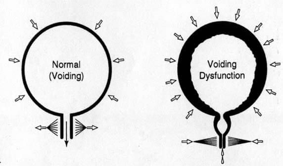
Urotherapy is a non-invasive treatment aimed at managing dysfunctional voiding and irregular bowel movement habits, which can affect urinary function and overall quality of life.
Dysfunctional Voiding
In dysfunctional voiding, the urethral sphincter fails to relax when the bladder contracts, making urination difficult and increasing bladder pressure. This can lead to urinary tract infections.
Irregular Bowel Movements
Irregular bowel movements can impact the bladder by increasing pressure on it, causing pelvic floor spasms that complicate urination. Proper management of both bladder and bowel function is essential for overall pelvic health.
Why Urotherapy is Necessary
Urotherapy focuses on normalizing bladder and bowel functions to improve quality of life. Treatment involves bladder training, pelvic muscle relaxation, and the use of medications if necessary.
Key Components of Urotherapy
Therapy may include recommendations on fluid intake, timed urination, and bowel management, along with dietary adjustments like increasing fiber and ensuring daily, scheduled bowel movements.
Who Provides Urotherapy?
Urotherapy is typically provided by a doctor or trained nurse counselor, who will assess the patient's condition and offer tailored recommendations.
What to Expect Next
After initial consultations, patients are encouraged to track their bladder activity through a bladder diary, which helps guide further treatment and track improvements.
Varicocele
A varicocele is an abnormal enlargement of the veins within the spermatic cord, often detected as a painless scrotal swelling. Some may experience discomfort or a "dragging" sensation. When standing, the veins enlarge; when lying down, they shrink.
More common in males after puberty, varicoceles are found in roughly 16% of adolescent boys and typically affect the left side. Though often asymptomatic, varicoceles are linked to infertility, as they can impair sperm production due to increased scrotal temperature.
If a varicocele causes pain or if the testicle appears underdeveloped, surgical treatment may be necessary. Microsurgical subinguinal repair is generally considered the least invasive method, using an operating microscope to ligate the dilated veins.
Vesicoureteral Reflux (VUR) in Children
Understanding VUR:
If your child has been diagnosed with Vesicoureteral Reflux (VUR), there are effective treatments available. Here's what VUR is and how it's typically managed.
What is VUR?
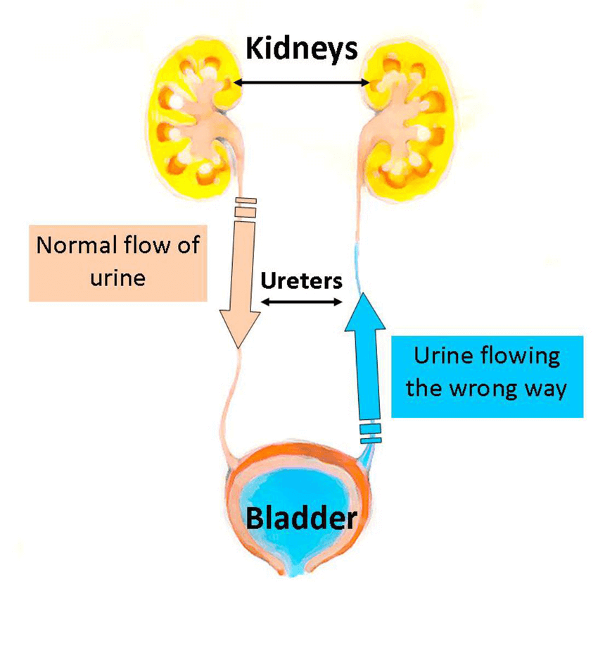
Vesicoureteral Reflux (VUR) is a condition where urine flows backward from the bladder into the kidneys. Normally, urine flows only one way, from the kidneys through the ureters to the bladder. VUR can occur on one or both sides of the urinary tract.
What Causes VUR?
VUR occurs in about 1% of children. It may be caused by a defect in the valve mechanism where the ureter meets the bladder (primary reflux) or due to other issues in the bladder or urinary tract (secondary reflux). Secondary reflux resolves once the underlying issue is treated. In older children, factors such as infrequent urination or constipation may contribute to VUR.
Is VUR Serious?
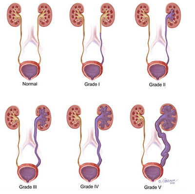
While VUR itself is typically not severe, it is important to treat it due to its potential link to urinary infections, kidney scarring, and possible renal failure. The severity of VUR is graded from I (mild) to V (severe), with higher grades requiring more aggressive treatment.
Testing for VUR
Tests to evaluate VUR typically include ultrasound, VCUG (voiding cystourethrogram), and DMSA (Dimercapto Succinic Acid) kidney scans to assess kidney scarring. A family history of VUR increases the likelihood of the condition in children, so it is crucial to discuss testing with your healthcare provider.
Who Should Be Tested?
Any child with urinary tract infections (UTIs) should undergo testing for VUR. The specific tests performed will depend on the child's age, symptoms, and ultrasound results.
Note that ultrasound alone cannot reliably detect VUR.
Why Should VUR Be Treated?
Treating VUR is vital to prevent kidney damage and protect the urinary system. Treatment involves managing infections and correcting the reflux.
How Do I Know if My Child Has VUR?
Symptoms of a urinary tract infection include:
- Foul-smelling or cloudy urine
- Fever
- Abdominal pain
- Burning or pain during urination
- Frequent and urgent urination
- Urinary tract infections
VUR itself does not typically show symptoms, but associated problems such as urinary incontinence or frequent infections may indicate its presence. Infants may exhibit non-specific symptoms like diarrhea or fever.
Treatment Options for VUR
There are three main treatment options for VUR:
- Antibiotics – Used to prevent infections while low-grade VUR resolves naturally.
- Surgery – Corrects high-grade VUR by fixing the ureters and often cures the condition.
- Endoscopic Treatment – Involves injecting a substance where the ureter meets the bladder in low-grade VUR.
How Do I Decide the Right Treatment for My Child?
Work with your healthcare provider to weigh the options together. Considerations include the severity of VUR, other medical conditions, and family preferences. Surgery may be recommended for severe cases, while antibiotics or endoscopic treatment may be more suitable for lower grades.
Follow-Up Care
Regular follow-up visits are essential to monitor the child's progress and ensure the chosen treatment is effective. Discuss the follow-up protocol with your doctor.
Normal Urine Flow
VESICOURETERAL REFLUX IN CHILDREN (VUR)